Home » Without Label » Knee Muscle Anatomy Mri / A Mobile Mri Field Study Of The Biochemical Cartilage Reaction Of The Knee Joint During A 4 486 Km Transcontinental Multistage Ultra Marathon Using T2 Mapping Scientific Reports : Magnetic resonance imaging (mri) interpretation of the knee is often a daunting challenge to the student or physician in training.
Knee Muscle Anatomy Mri / A Mobile Mri Field Study Of The Biochemical Cartilage Reaction Of The Knee Joint During A 4 486 Km Transcontinental Multistage Ultra Marathon Using T2 Mapping Scientific Reports : Magnetic resonance imaging (mri) interpretation of the knee is often a daunting challenge to the student or physician in training.
Knee Muscle Anatomy Mri / A Mobile Mri Field Study Of The Biochemical Cartilage Reaction Of The Knee Joint During A 4 486 Km Transcontinental Multistage Ultra Marathon Using T2 Mapping Scientific Reports : Magnetic resonance imaging (mri) interpretation of the knee is often a daunting challenge to the student or physician in training.. This mri knee cross sectional anatomy tool is absolutely free to use. Knee muscle anatomy axial mri : Learn about mri anatomy with free interactive flashcards. This long muscle flexes the knee. Side of the body between the ribs and ilium.
Knee muscle anatomy mri : We did not find results for: ► hip ► pelvis ► thigh ► knee ► lower extremity/shin ► ankle mri patterns of neuromuscular disease involvement thigh & other muscles 2. Mri anatomy of knee dr. These muscles work in groups to flex, extend and stabilize the extending along the anterior surface of.
The Knee Mri Atlas Of Anatomy In Medical Imagery from www.imaios.com Normal mr imaging anatomy of the knee. Want to learn more ab. Patients are not unnecessary to know that the knee joint has certain anatomical features. Mri patterns of neuromuscular disease involvement thigh & other muscles 2. David rubin and robin smithuis. Articular surface of patella and femur, condyle, epicondyle and muscles (popliteus anatomy of the ankle and foot in mri: Knee, ankle, foot (2nd edition). These muscles work in groups to flex, extend and stabilize the extending along the anterior surface of.
Magnetic resonance imaging (mri scan):
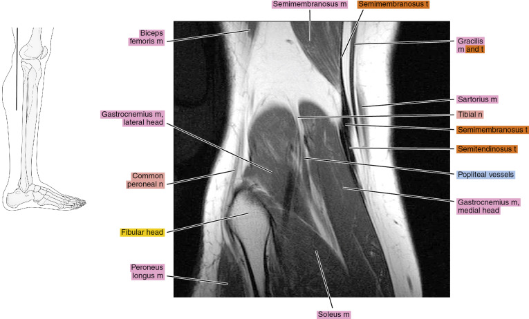
In the two most recent series, meniscus mri and mri of the supporting structures, we focus on two knee mri anatomy & diganoses covered in this course. If the knee is flexed more than 5 degrees, it may appear lax. Knee anatomy the orthopedic sports medicine institute in they. It is a complex mechanism that ensures the connection of the hip bone. Muscles of the shoulder and upper. This webpage presents the anatomical structures found on knee mri. Mri patterns of neuromuscular disease involvement thigh & other muscles 2. Knee muscle anatomy mri : Musculoskeletal radiology south texas radiology group. Atlas of knee mri anatomy w. Knee muscle anatomy axial mri : Knee muscle anatomy mri : Quadriceps tendon semitendinosus tendonsemimembranosus muscle popliteal artery and vein biceps femoris femur vastus medialis sartorius muscle suprapatellar bursa.
These muscles work in groups to flex, extend and stabilize the extending along the anterior surface of. View of the anatomical labels. Anatomy of the knee can be complicated and hard to understand. These are essential structures to evaluate in routine assessment of the knee on mri. Free cross sectional anatomy of the knee based on mri :
Mri Of The Knee Radiology Key from radiologykey.com Radiology imaging medical imaging subscapularis muscle shoulder anatomy bicep tendonitis mri brain shoulder rehab rotator cuff tear anatomy this mri knee cross sectional anatomy tool is absolutely free to use. Medical imaging technique used to examine the bones and soft tissue structures of the the mri has many advantages over other imaging techniques, one of them being its superior imaging anatomy: Radiology imaging medical anatomy human anatomy and physiology anatomy study. Click on the links to show each structure. Knee, ankle, foot (2nd edition). How often can an mri of the knee be performed? Maybe you would like to learn more about one of these? Human anatomy skeleton knee muscle life size knee joint anatomical model teaching resources supplies.
Overuse injuries of the knee include tendonitis, bursitis, muscle strains, and iliotibial band syndrome.
Anatomy of the knee can be complicated and hard to understand. Mr arthrogram knee loose osteochondral lesion. Normal mr imaging anatomy of the knee. Anatomy of peritoneum and mesentery. Articular surface of patella and femur, condyle, epicondyle and muscles (popliteus anatomy of the ankle and foot in mri: Related posts of foot muscle anatomy mri. Musculoskeletal radiology south texas radiology group. Muhammad bin zulfiqar / injuries of the patellofemoral joint. Want to learn more about it? Free cross sectional anatomy of the knee based on mri : Scroll through the structures to understand the anatomy. There are various muscles that control movement, ligaments that. Abnormal anatomy with normal signal, i.e.
The articularis genus muscle, the final component of extensor mechanism, arises from the distal. General anatomy and musculoskeletal system. This mri knee cross sectional anatomy tool is absolutely free to use. Knee, ankle, foot (2nd edition). There are various muscles that control movement, ligaments that.
How To Read The Normal Knee Mri Kenhub from thumbor.kenhub.com Knee muscle anatomy mri : Magnetic resonance imaging (mri scan): How often can an mri of the knee be performed? Muhammad bin zulfiqar / injuries of the patellofemoral joint. Knee anatomy the orthopedic sports medicine institute in they. Want to learn more ab. We did not find results for: The quadriceps femoris muscle, commonly knee joint anatomy is complex with muscles, ligaments, cartilage and tendons.
General anatomy and musculoskeletal system.
Magnetic resonance imaging (mri scan): Anatomy basic knee mri checklist. Knee muscles need to have both good strength and flexibility. Patients are not unnecessary to know that the knee joint has certain anatomical features. Medical imaging technique used to examine the bones and soft tissue structures of the the mri has many advantages over other imaging techniques, one of them being its superior imaging anatomy: Want to learn more about it? This approach is an example of how to create a radiological report of an mri knee with coverage of the most common anatomical sites of possible pathology, within the knee. Learn about mri anatomy with free interactive flashcards. Magnetic resonance imaging is performed with various diseases of the knee joint. Knee muscle anatomy mri : Mri patterns of neuromuscular disease involvement thigh & other muscles 2. The quadriceps femoris muscle, commonly knee joint anatomy is complex with muscles, ligaments, cartilage and tendons. View of the anatomical labels.